Labelled Radius Bone / Upper Limbs Mistryland. These bones are specially designed in order to enable the movements that are unique for the upper limb, such are supination and pronation. The ulna is on the medial side of the forearm and forms a hinge joint with the humerus at the elbow. #labelled diagram of radius bone. Introduction to the radius and ulna bones anatomy the radius and ulna are the bones of the forearm. The humerus (/ ˈhjuːmərəs /, plural:
It extends from the lateral side of the elbow to the thumb side of the wrist and runs parallel to the ulna. In the classical anatomical position, the radius is found laterally, while the ulna is the medial of the two bones. There are 30 bones in each upper limb. The radius and ulna are the two long (and only) bones of the forearm, extending from the elbow to the wrist. The radius bone is homologous to the medial bone of the leg, tibia.
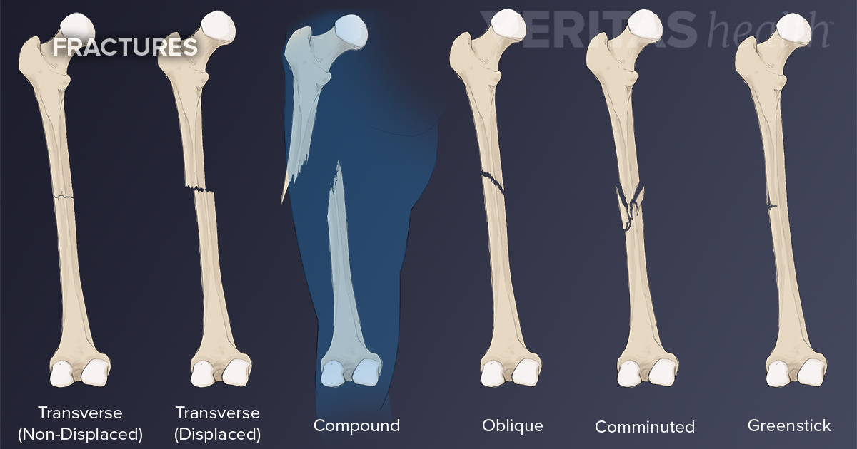
Bone Break Vs Fracture from embed.widencdn.net Introduction to the radius and ulna bones anatomy the radius and ulna are the bones of the forearm. In concert with each other, the two bones play a vital role in how the forearm rotates. This article explains the bone structure of the human body, using a labeled skeletal system diagram and a simple technique to memorize the names of all the bones. It is one of the two bones of the forearm, the other being the ulna. The radius bone is the lateral bone of the forearm, and is homologous with the tibia of the lower limb. Bone labeling radius and ulna. The ulna is usually slightly longer than the radius, but the radius is thicker. What is the radial bone facts, where is the radius located in arm, what does it do, anatomy (type, parts, joints formed), labeled diagram.
Prismatic in shape, it starts from the lateral side of the elbow and continues to the thumb side of the wrist.
What is the radial bone facts, where is the radius located in arm, what does it do, anatomy (type, parts, joints formed), labeled diagram. The radius bone is a long horizontal bone present in the forearm and is also called the radial bone. The radius or radial bone is one of the two large bones of the forearm, the other being the ulna. This is the head, and it has a depression at the top that forms a joint with the capitulum of the humerus bone. 1 2 interosseous border of the radius (margo interosseus radii) is the medial edge (margin) of the bone where the interosseous membrane attaches. The radius bone is a long horizontal bone present in the forearm and is also called the radial bone. The forearm is the region of the upper limb that extends from the elbow to the wrist. The ulna is on the medial side of the forearm and forms a hinge joint with the humerus at the elbow. Related posts of labelled diagram of radius bone muscles and bones in the arm. There are 30 bones in each upper limb. Carpal bones on xray 12 photos of the carpal bones on xray carpal bone dislocation x ray, carpal bone fracture x ray, carpal bones. Related posts of labelled diagram of radius bone bones in the foot diagram. Correctly label the following bones and anatomical features of the inferior view of the skull.
1 2 interosseous border of the radius (margo interosseus radii) is the medial edge (margin) of the bone where the interosseous membrane attaches. On the upper part of the shaft is a rough projection, the radial tuberosity, which receives the biceps tendon.a ridge, the interosseous border, extends the length of the shaft and provides attachment for the interosseous membrane. The radius and ulna are the two bones of the forearm. Related posts of labelled diagram of radius bone muscles and bones in the arm. The radius bone is a long horizontal bone present in the forearm and is also called the radial bone.
8 2 Bones Of The Upper Limb Anatomy Physiology from open.oregonstate.education The radius bone is a long horizontal bone present in the forearm and is also called the radial bone. Humeri) is a long bone in the arm that runs from the shoulder to the elbow. The base of the hand contains eight bones, each called a carpal bone, and the palm of the hand is formed by five bones, each called a metacarpal bone. The forearm is the region of the upper limb that extends from the elbow to the wrist. Palatine process of maxilla, sphenoid bone, temporal bone, occipital condyle, zygomatic bone, vomer, mandibular fossa, styloid process. The radius bone is homologous to the medial bone of the leg, tibia. These bones are specially designed in order to enable the movements that are unique for the upper limb, such are supination and pronation. The radius bone is a long horizontal bone present in the forearm and is also called the radial bone.
A basic human skeleton is studied in schools with a simple diagram.
Prismatic in shape, it starts from the lateral side of the elbow and continues to the thumb side of the wrist. The radius or radial bone is one of the two large bones of the forearm, the other being the ulna. It is a long bone that runs parallel to the radius, along the forearm. 1 2 interosseous border of the radius (margo interosseus radii) is the medial edge (margin) of the bone where the interosseous membrane attaches. Muscles and bones in the arm 12 photos of the muscles and bones in the arm muscles. #labelled diagram of radius bone. It is not to be confused with humoral immunity. There are 30 bones in each upper limb. The humerus is the single bone of the upper arm, and the ulna (medially) and the radius (laterally) are the paired bones of the forearm. The radius bone (os radius) supports the lateral (thumb) side of the forearm and the ulna bone (os ulna) supports the medial (little finger) side. Therefore the radius is considered to be the larger of the two. The radius bone is a long horizontal bone present in the forearm and is also called the radial bone. The radius is a long bone in the forearm.
What this does is it stabilizes the joint and it allows the radius to rotate against the radial notch on the ulna and also at. Its upper concave surface articulates with the humerus (upper arm bone) above, and the side surface articulates with the ulna. In concert with each other, the two bones play a vital role in how the forearm rotates. The radius bone is a long horizontal bone present in the forearm and is also called the radial bone. The radius bone is the lateral bone of the forearm, and is homologous with the tibia of the lower limb.
Radius Bone Artwork Stock Image C020 9125 Science Photo Library from media.sciencephoto.com #labelled diagram of radius bone. #labelled diagram of radius bone. Where is the radius bone located in the arm it is located on the thumb side of the hand, lying laterally in the lower arm, parallel in reference to the ulna 1, 2. The ulna is usually slightly longer than the radius, but the radius is thicker. Bone growth in the radius of the styloid process. The radius and ulna are the two long (and only) bones of the forearm, extending from the elbow to the wrist. So, the scaphoid being the first bone in the proximal row means it articulates with the radius. The radius articulates in four places:
It is a long bone that runs parallel to the radius, along the forearm.
The radius was a bone in the humanoid skeletal system. The humerus (/ ˈhjuːmərəs /, plural: These bones are specially designed in order to enable the movements that are unique for the upper limb, such are supination and pronation. Palatine process of maxilla, sphenoid bone, temporal bone, occipital condyle, zygomatic bone, vomer, mandibular fossa, styloid process. The ulna is on the medial side of the forearm and forms a hinge joint with the humerus at the elbow. The humerus is the single bone of the upper arm, and the ulna (medially) and the radius (laterally) are the paired bones of the forearm. The lower arm bones form the wrist joint with the carpals, a group of eight small bones that give added flexibility to. The radius and ulna are the two bones of the forearm. It extends from the lateral side of the elbow to the thumb side of the wrist and runs parallel to the ulna. Therefore, the forearm is in the. Carpal bones on xray 12 photos of the carpal bones on xray carpal bone dislocation x ray, carpal bone fracture x ray, carpal bones. You will be required to label the ulnar notch, styloid process of ulna, trochlear notch, proximal radioulnar joint, olecranon process, coronoid process, distal radioulnar joint, etc. It is instrumental in the shaping and use of hands.
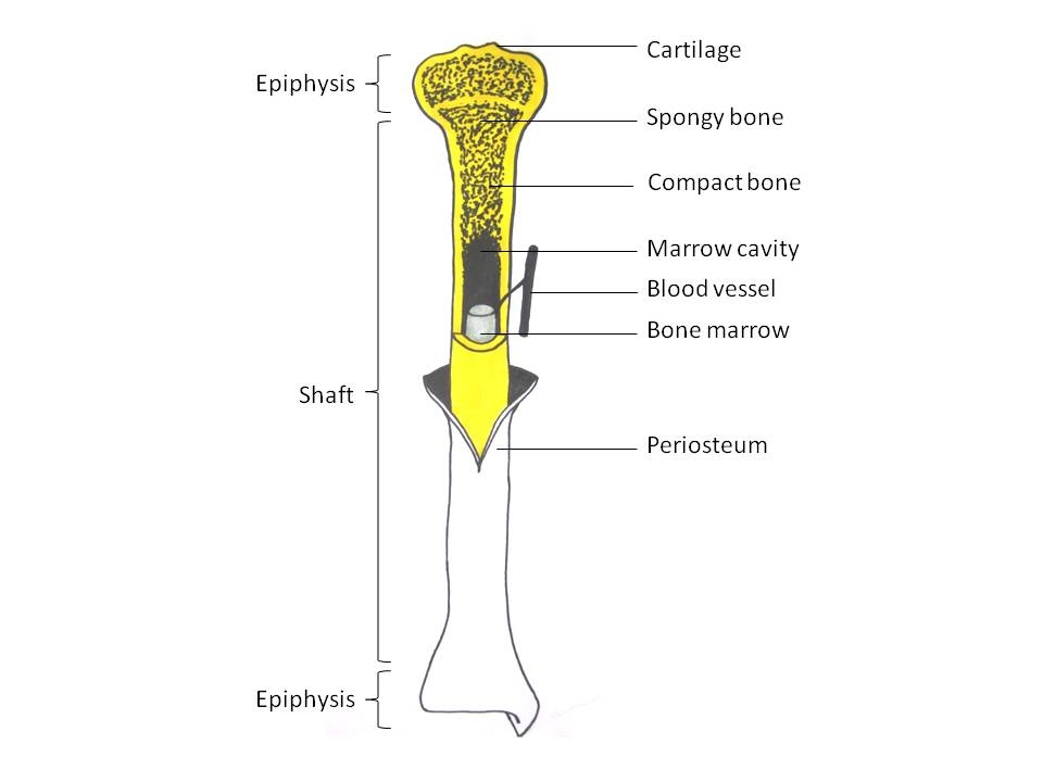
Long Bone With Diagram : Bone Structure And The Anatomy Of Long Bones. Long bones have a thick outside layer of compact bone and an inner medullary cavity containing bone marrow. A long bone has a shaft and 2 ends. Long bone with diagram : The diaphysis and the epiphysis. The bone on the left in the image is the :
• all bones of the limbs, except the patella, wrist and ankle bones, are long bones. Game statistics long bone diagram from www.purposegames.com they include fill in the blank anatomy diagrams, completed diagrams and numbered quizzes. Parts of long bone (applies to other bones too). The diaphysis and the epiphysis. It is a more structured approach than some other tools available for brainstorming causes
Parts Of A Long Bone 2 2 2 Human Skeleton By Openstax Page 4 4 Jobilize from www.jobilize.com What is label number 4 pointing to in the diagram? Long bone shaft anatomy system human body anatomy diagram and. They are one of five types of bones: A long bone has two parts: The structure of a long bone allows for the best visualization of all of the parts of a bone figure 1. Smartdraw includes 1000s of professional healthcare and anatomy chart templates that you can modify and make your own. The diaphysis is the tubular shaft that runs between the proximal and distal ends of the bone. November 14, 2017november 14, 2017 / clarebosanko.
Long bones have a thick outside layer of compact bone and an inner medullary cavity containing bone marrow.
A long bone has two parts: They are one of five types of bones: The second metatarsal bone is the longest. Parts of long bone (applies to other bones too). Label number 3 in the diagram is pointing to : In a long bone, for example, at about 6 to 8 weeks after conception, some of the mesenchymal cells differentiate into chondroblasts (cartilage cells) that form the hyaline cartilaginous skeletal precursor of the bones (figure 6.4.2a). A fishbone diagram is a visual way to look at cause and effect. Long bones of the leg include the femur, tibia, fibula, metatarsals, and phalanges. It is the only bone making up the upper arm. Humerus bone quiz anterior markings : Long bones in the arm include the humerus, radius, ulna, metacarpals, and phalanges. The structure of a long bone allows for the best visualization of all of the parts of a bone (figure 1). The diaphysis is the tubular shaft that runs between the proximal and distal ends of the bone.
Long bones of the leg include the femur, tibia, fibula, metatarsals, and phalanges. The structure of a long bone allows for the best visualization of all of the parts of a bone (figure 1). Long bones are one of the five bone types that are classified by shape. Label number 3 in the diagram is pointing to : A typical long bone shows the gross anatomical characteristics of bone.
Blood Supply Of Long Bone Primary Category Anatomy Qa from i0.wp.com A fishbone diagram is a visual way to look at cause and effect. There is a printable worksheet available for download here so you can take the quiz with pen and paper. Humerus bone quiz anterior markings : Students fill in the boxes with the names of the bones. There is a printable worksheet available for download here so you can take the quiz with pen and paper. A long bone is a bone that has greater length than width. The blood vessels inside a bone. This diagram depicts final long bone diagram.human anatomy diagrams show internal organs, cells, systems, conditions, symptoms and sickness information and/or tips for healthy living.
What do we mean by an 'articulation'?
Smartdraw includes 1000s of professional healthcare and anatomy chart templates that you can modify and make your own. Long bones are one of the five bone types that are classified by shape. There is a printable worksheet available for download here so you can take the quiz with pen and paper. The bone on the right in the image is the : What do we mean by an 'articulation'? Long bones lengthen at the epiphyseal plate with the addition of bone tissue and increase in width by a process called appositional growth. The structure of a long bone allows for the best visualization of all of the parts of a bone (figure 6.7). The skeleton of the arms and legs are made up of mostly long bones. Long bones in the arm include the humerus, radius, ulna, metacarpals, and phalanges. The structure of a long bone allows for the best visualization of all of the parts of a bone ((figure)). A cause and effect diagram, often called a fishbone diagram, can help in brainstorming to identify possible causes of a problem and in sorting ideas into useful categories. The diaphysis is the tubular shaft that runs between the proximal and distal ends of the bone. A long bone has two parts:
Bones at the base of the skull and long bones form via endochondral ossification. In this video we discuss the parts of a long bone and some of the functions of each of those bone parts. Choose from 500 different sets of long bone diagram flashcards on quizlet. Used figure 6.2 in book. This diagram depicts final long bone diagram.human anatomy diagrams show internal organs, cells, systems, conditions, symptoms and sickness information and/or tips for healthy living.
Long Bone Wikipedia from upload.wikimedia.org A typical long bone shows the gross anatomical characteristics of bone. Long bone diagram labeled find out more about long bone diagram labeled. The femur, or thighbone, is the longest and largest bone in the human body. Humerus bone quiz anterior markings : Some descriptions for confusing parts.omit number 13 in the picture. You need to get 100% to score the 10 points available. The structure of a long bone allows for the best visualization of all of the parts of a bone ((figure)). A 'crest' on a bone is :
The diaphysis is the tubular shaft that runs between the proximal and distal ends of the bone.
Long bones are one of the five bone types that are classified by shape. Students fill in the boxes with the names of the bones. A fishbone diagram is a visual way to look at cause and effect. A typical long bone shows the gross anatomical characteristics of bone. Parts of long bone (applies to other bones too). Spongy bone, also known as cancellous bone or trabecular bone, is a very porous type of bone. November 14, 2017november 14, 2017 / clarebosanko. Long bones include the humerus (upper arm), radius (forearm), ulna (forearm), femur (thigh), fibula (thin bone of the lower leg), tibia (shin bone) , phalanges (digital bones in the hands and feet), metacarpals (long bones within the hand), and metatarsals (long bones. The end of a long bone. Long bones lengthen at the epiphyseal plate with the addition of bone tissue and increase in width by a process called appositional growth. Press enter to view all search results. You need to get 100% to score the 10 points available. The long bones are those that are longer than they are wide.